
Understanding MRD in AML and beyond through the lens of single-cell multiomics.
Quick answer: Measurable residual disease (MRD), also called minimal residual disease, is the small population of malignant cells that survives cancer treatment below the limit of conventional detection. MRD status is one of the strongest predictors of relapse and survival across hematologic malignancies such as AML, ALL, multiple myeloma, and CLL. Most MRD tests answer only whether residual disease is present. Single-cell multiomics goes further, resolving which clones persisted, why they survived, and how they drive relapse.
Measurable, or minimal, residual disease has become one of the most consequential biomarkers in oncology. A growing body of evidence ties MRD status to relapse risk, depth of treatment response, and overall survival, and regulators are increasingly willing to treat it as an early efficacy endpoint. In acute myeloid leukemia (AML), MRD-negative patients show roughly double the five-year overall survival of MRD-positive patients after intensive induction (Short et al., 2020).
Yet most MRD technologies answer a single question: is disease present? That binary readout is clinically useful but biologically thin. Two patients with identical MRD levels can carry very different clonal architectures and face very different relapse trajectories. Emerging single-cell approaches reframe the question entirely, asking which clones survived therapy, what genotype-to-phenotype features let them persist, and what is likely to happen next. This piece uses AML as the primary example because it is the disease where clonal complexity is best characterized, but the argument extends to ALL, multiple myeloma, CLL, and, increasingly, solid tumors.
What is Measurable residual disease (MRD)?
Measurable residual disease (MRD) is the small number of cancer cells that persist after treatment, below the threshold of standard morphologic or molecular detection.
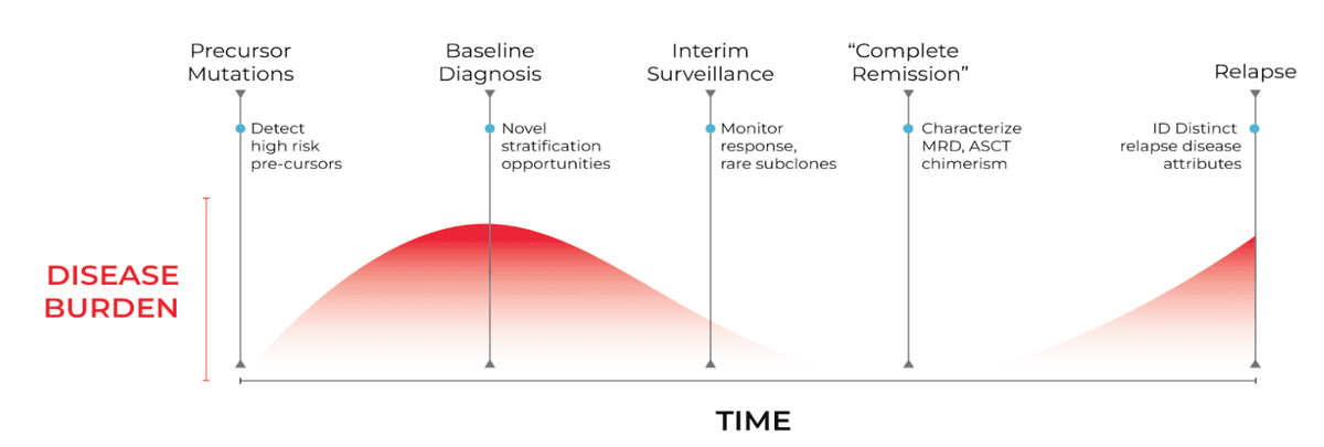
MRD describes the small numbers of malignant cells that remain after treatment has produced an apparent complete remission. These cells sit below the threshold of morphologic assessment, which typically detects disease only above 5% blasts, and often below the floor of routine molecular workups. Their presence, even at very low frequency, signals that the disease has not been eradicated.
MRD matters because it is one of the strongest available predictors of relapse. It serves four overlapping roles in practice:
- as a predictor of relapse
- as a tool for post-remission risk stratification
- as a means of longitudinal treatment monitoring
- as an endpoint in clinical trials.
The European LeukemiaNet recommends a 0.1% threshold for MRD negativity by multiparameter flow cytometry and defines complete remission (CR) without MRD (CRMRD−) as the optimal AML response (ELN MRD recommendations, Heuser et al.).
Clinical and regulatory interest has increased in parallel to each other. MRD is now evaluated routinely in AML, acute lymphoblastic leukemia (ALL), multiple myeloma, chronic lymphocytic leukemia (CLL), and mantle cell lymphoma, with emerging applications in solid tumors via circulating tumor DNA, also referred to as ctDNA. As therapy intensifies and remission rates rise, the clinically meaningful question shifts from whether a patient entered remission to how deep and durable that remission really is.
How is MRD measured? Current technologies for MRD testing
MRD is measured primarily by multiparameter flow cytometry, PCR/ddPCR, and bulk next-generation sequencing, with single-cell multiomics (DNA sequencing + immunophenotyping) emerging as a higher-resolution option.
Three main methods dominate MRD assessment today, and each makes a different compromise between sensitivity, accessibility, and biological resolution.
Multiparameter flow cytometry is established, widely available, and fast, returning results in hours from fresh marrow. It tracks leukemia-associated immunophenotypes and the difference-from-normal pattern. Its limitations are well known. Immunophenotypic shift between diagnosis and relapse can move the target, interpretation is operator-dependent and expertise-limited, and it returns essentially no genomic information about the surviving cells which is detrimental because not all cancer cells will have a phenotype that is different from the wildtype.
Bulk next-generation sequencing offers high sensitivity, with unique multiplier indicator-based error correction pushing variant allele frequency (VAF) detection toward 10⁻⁴ to 10⁻⁶, and it can track patient-specific molecular markers longitudinally. But bulk NGS reports population averages. A VAF is a property of a sample, not a cell, so the assay cannot tell whether two mutations sit in the same cell or in distinct clones therefore it cannot reconstruct clonal architecture which makes it challenging to track mutant clonal evolution.
PCR-based methods, including RQ-PCR and digital droplet PCR, have good sensitivity for defined targets such as NPM1 or fusion transcripts, reaching 10⁻⁵ or better. Their constraint is they detect only known, pre-specified targets and provide no view of mutant clones that lack the tracked marker.
The shared takeaway is that current technologies can often detect residual disease but struggle to characterize it. They tell you a signal is there without telling you what the surviving cell actually is.
Table 1 — Comparison of MRD assessment modalities by sensitivity and biological resolution.
| Method | Sensitivity | Molecular Resolution | Clonal Resolution |
|---|---|---|---|
Multiparameter flow | Moderate (~0.1%) | Low | Low |
PCR / ddPCR | High (targeted) | Targeted | Low |
Bulk NGS | High | Moderate | Low |
Single-cell multiomics | High | High | High |
Why is MRD in AML so difficult to interpret?
MRD in acute myeloid leukemia (AML) is hard to interpret because AML is genetically heterogeneous because the residual disease is a mix of mutant and wildtype clones, and bulk tests cannot tell which surviving clones will drive relapse.
AML is the clearest illustration of why detection alone is insufficient because it is among the most genetically heterogeneous hematologic malignancies. A single patient typically harbors a founder clone, multiple subclones, and minor populations carrying resistance-associated lesions. Induction chemotherapy and targeted agents frequently eliminate the dominant clone while sparing resistant subclones that then seed relapse.
This is precisely where bulk sequencing methods fail in characterizing the disease. Suppose bulk sequencing of a remission marrow reports mutation A at 1% VAF and mutation B at 2% VAF. The data cannot identify if A and B are in the same cell, marking a single double-mutant clone, or in separate cells, marking two independent populations, and if either one is or can be an emerging resistant clone? Co-occurrence and mutual exclusivity are invisible to a population average seen with bulk sequencing.
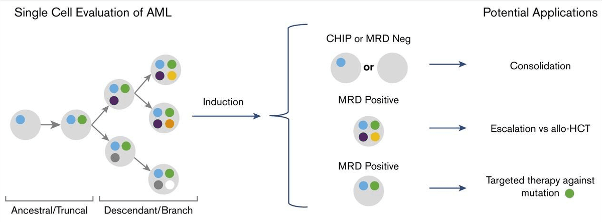
This means the clinical questions stack up quickly. Which clones survived induction? Which clones are most likely to drive relapse? Are pre-leukemic clones persisting, and is clonal hematopoiesis being mistaken for true MRD? That last question should be taken seriously. Mutations in DNMT3A, TET2, and ASXL1 (the “DTA” genes) often persist through remission as clonal hematopoiesis of indeterminate potential (CHIP) without conferring worse outcomes, whereas persistence of non-DTA mutations carries clear prognostic weight (Jongen-Lavrencic / molecular MRD analyses). Many MRD pipelines therefore exclude DTA mutations but doing so on a bulk VAF alone is a blunt instrument.
Recent work shows the deeper problem that persistent post-remission clonal hematopoiesis actively shapes relapse trajectories, and the dominant clone at MRD assessment is not always the clone that dominates at relapse (Ediriwickrema et al., single-cell AML MRD). This is why clonal context matters. MRD is not simply a number, where two patients with identical MRD percentages may face very different relapse risks depending on which clones make up that residual signal and how those clones evolve within the population, eventually causing relapse.
How does single-cell improve MRD analysis?
Unlike bulk sequencing which averages cellular data, single-cell multiomics genotypes cells individually to precisely map clonal architecture, zygosity, and mutational co-occurrence. Crucially, it links these genetic profiles directly to cell-surface protein phenotypes, allowing clinicians to identify targetable receptors on rare MRD clones and distinguish them from benign mutations.
Single-cell DNA sequencing resolves the ambiguity that defeats bulk methods by reading genotypes one cell at a time. Instead of asking what fraction of alleles carry a mutation, it asks which mutations co-occur within the same cell which aids in the reconstruction of true clonal architecture. For diseases like AML, matching single-cell genotypes to cell phenotypes directly links mutation to function, determining if a rare clone is benign or capable of evolving therapy resistance.In a head-to-head analysis, single-cell mutational profiling identified MRD in 40% of remission samples versus 9% by flow cytometry, and detected clonal mutations in remission marrows from 8 of 10 patients who later relapsed (Blood Advances, 2020).
Four capabilities of single-cell MRD assays include,
- Clonal architecture: the assay identifies which specific clones survived treatment and how they relate to the diagnostic clone.
- Evolutionary trajectories: longitudinal single-cell sampling shows how relapse-driving populations emerge and expand.
- Genotype-to-phenotype analysis: genotype-to-phenotype assessment is performed by profiling genetic mutations and linking them directly to the surface receptors detected through immunophenotyping on the same cell, so a specific clone's mutations can be mapped to its surface-marker profile and cell state rather than inferred from separate bulk DNA sequencing and flow cytometry measurements.
- Rare mutant detection: clinically relevant subpopulations hidden inside a bulk averaged data become visible as discrete mutant clones.
This is where the Tapestri platform from Mission Bio solves the current problem of MRD assays. Its two-step droplet workflow encapsulates single cells, lyses them, and barcodes genomic DNA so that thousands of cells are genotyped individually across a targeted panel of recurrently mutated AML genes. The Tapestri single-cell MRD (scMRD) assay for AML pairs a curated AML DNA panel and surface-protein panel, integrating mutational and immunophenotypic signatures in one cell to separate true leukemic clones from CHIP.
Where else is MRD used beyond AML?
MRD is used to monitor treatment and predict relapse across ALL, multiple myeloma, and CLL, with emerging ctDNA-based applications in solid tumors.
The same logic applies wherever residual disease is clonally heterogeneous. In acute lymphocytic leukemia (ALL), MRD is already a backbone of therapy monitoring and relapse prediction, and single-cell resolution helps distinguish lineage-switching and resistant subclones that complicate immunophenotypic tracking. In multiple myeloma (MM), spatial heterogeneity and resistant subclones challenge any single-site bulk sequencing measurement, and MRD is increasingly used as a trial endpoint (discussed below).
In chronic lymphocytic leukemia (CLL), the relevant biology is clonal persistence and the evolution of resistance under targeted agents such as BTK and BCL2 inhibitors, where understanding which subclone survives is more informative than a bulk burden estimate. In solid tumors, MRD is more nascent where circulating tumor cells and ctDNA-based liquid biopsy offer a route to monitoring targeted-therapy response, and single-cell characterization of rare disseminated cells is a forward-looking opportunity rather than established practice. It is best framed as where the field is heading, not where it already is.
Is MRD a valid endpoint in clinical trials?
Yes. MRD is increasingly accepted as an early surrogate endpoint. In April 2024 the FDA's ODAC voted that MRD-negative complete response is reasonably likely to predict clinical benefit in multiple myeloma.
For biotech and pharma audiences, the endpoint story is the most immediately relevant. MRD is increasingly used as a surrogate endpoint, an early efficacy signal, and a stratification tool that can shorten trials otherwise gated on progression-free or overall survival. In April 2024, the FDA’s Oncologic Drugs Advisory Committee (ODAC) voted that MRD-negative complete response is reasonably likely to predict clinical benefit in multiple myeloma, and a January 2026 draft guidance now consolidates how developers can use it (MRD roadmap, Blood Cancer Discovery, 2025; FDA Draft Guidance: MRD in Multiple Myeloma. Jan 2026.).
But a falling MRD rate is a coarse signal of mechanism. Drug developers increasingly want to know not just whether MRD decreased, but which clones persisted, which mutations escaped, and what resistance mechanisms emerged under selective pressure. Single-cell approaches provide exactly that biological resolution, turning an MRD endpoint into a window on how a therapy reshapes the clonal landscape which is information that informs combination therapeutic strategy and patient selection long before survival data matures.
What is the future of MRD testing?
The future of MRD testing is single-cell multiomic and longitudinal monitoring where combining DNA, RNA, and protein at single-cell resolution explains why residual cells persist, not just whether they are present.
The trajectory points toward richer, more integrated MRD assessment, with AML emerging as the clear next focus for clinical endpoint validation. Single-cell multiomic profiling that layers targeted DNA genotyping, same-cell surface-protein immunophenotyping, and targeted transcriptional readout will tie surviving genotypes (FLT3-ITD, NPM1, IDH1/2, RAS-pathway and splicing-factor lesions) to the functional cell states that let them evade therapies, and will separate residual leukemic clones from DTA-driven clonal hematopoiesis with far more confidence than a bulk VAF threshold allows. Longitudinal single-cell tracking across induction, consolidation, and post-transplant timepoints will resolve clonal evolution and the outgrowth of resistant subclones as a continuous trajectory rather than isolated snapshots. Additionally, scaling datasets to thousands of cells per sample puts the spotlight on efficient data processing. Single-cell multiomic experts and machine-learning algorithms offer valuable assistance, optimizing the way computational pipelines cluster cells into clones, phase co-occurring mutations, and interpret sparse single-cell signals.
The Mission Bio Tapestri platform brings this single-cell multiomic future to the present day, providing a powerful single-cell AML MRD assay that maps the exact clonal architecture clinicians need to predict therapy resistance and personalize patient care.
Conclusion
For years, MRD assessment has focused on a single determination asking the question, is residual disease present? Current MRD assays have been ok but are not the best making the next wave of MRD technologies seeks to answer a deeper question, what biological mechanisms allow residual cells to survive and eventually drive relapse? As the need for precision oncology progresses, understanding the clonal composition, evolutionary dynamics, and functional properties of residual disease may become as important as detecting it. Single-cell multiomic approaches are helping researchers move beyond standard of care MRD detection methods and toward a more comprehensive understanding of disease persistence and treatment resistance, which is the difference between knowing a patient is MRD-positive and knowing what to do about it.
Frequently Asked Questions About MRD
What is the difference between minimal residual disease and measurable residual disease?
They are the same concept. “Measurable residual disease” is the preferred current term because it emphasizes what assays can quantify, while “minimal residual disease” is the older, widely recognized name. Both share the abbreviation MRD.
What does MRD-positive mean?
MRD-positive means residual cancer cells were detected after treatment, below the level of standard morphologic assessment. MRD positivity is associated with a higher risk of relapse and shorter survival. In AML, MRD-negative patients show roughly double the five-year overall survival of MRD-positive patients.
What is the detection threshold for MRD in AML?
The European LeukemiaNet defines MRD negativity by multiparameter flow cytometry at a 0.1% (10⁻³) threshold. Molecular methods such as PCR and error-corrected NGS can reach 10⁻⁴ to 10⁻⁶, and single-cell DNA sequencing achieves comparable sensitivity while adding clonal resolution.
Can clonal hematopoiesis (CHIP) be mistaken for MRD?
Yes. Persistent DNMT3A, TET2, and ASXL1 (DTA) mutations often reflect clonal hematopoiesis rather than true leukemic MRD, and many bulk sequencing methods average signals so you cannot distinguish which cells have which mutation. Single-cell sequencing helps separate pre-leukemic CHIP clones from genuine residual disease by resolving co-occurring mutations within individual cells.
Why is single-cell MRD better than bulk MRD testing?
Bulk MRD reports an average variant allele frequency (VAF) across a sample and cannot tell whether two mutations are in the same cell. Single-cell MRD genotypes individual cells, revealing clonal architecture, rare resistant subpopulations, and the surviving clones most likely to drive relapse.
How does single-cell genotype-to-phenotype mapping improve MRD analysis?
Single-cell genotype-to-phenotype mapping improves MRD analysis by simultaneously evaluating DNA mutations and cell-surface protein expressions within the exact same cell. This proteogenomic approach allows clinicians to link a clone's genetic identity directly to its functional behavior. In diseases like AML, this multiomic resolution reveals whether a rare, mutation-carrying clone expresses targetable surface receptors, allowing teams to distinguish benign mutations from aggressive, therapy-resistant cells and select the precise follow-up therapies to eliminate them.
Selected References
1. Short NJ, Zhou S, Fu C, et al. Prognostic and therapeutic implications of measurable residual disease in acute myeloid leukemia. J Hematol Oncol. 2021. https://jhoonline.biomedcentral.com/articles/10.1186/s13045-021-01148-5
2. Heuser M, Freeman SD, Ossenkoppele GJ, et al. Measurable residual disease detection in AML: current challenges and future directions (ELN MRD Working Party recommendations). https://pmc.ncbi.nlm.nih.gov/articles/PMC10968436/
3. Ediriwickrema A, Aleshin A, Reiter JG, et al. Single-cell mutational profiling enhances the clinical evaluation of AML MRD. Blood Adv. 2020;4(5):943–952. https://ashpublications.org/bloodadvances/article/4/5/943/452671/Single-cell-mutational-profiling-enhances-the
4. Ediriwickrema A, et al. Single-cell mutational profiling enhances the clinical evaluation of AML MRD (full text). PMC7065471. https://pmc.ncbi.nlm.nih.gov/articles/PMC7065471/
5. Exclusion of persistent mutations in splicing factor genes and IDH2 improves the prognostic power of molecular MRD assessment in AML. Haematologica. https://haematologica.org/article/view/haematol.2023.283510
6. Persistent post-remission clonal hematopoiesis shapes the relapse trajectories of acute myeloid leukemia. (2025). https://www.sciencedirect.com/science/article/pii/S2473952925001016
7. Pellegrino M, Sciambi A, Treusch S, et al. Clonal evolution and changes in two AML patients detected with a novel single-cell DNA sequencing platform. Sci Rep. 2019;9:11407. https://www.nature.com/articles/s41598-019-47297-z
8. Multi-omic single-cell sequencing reveals discordant MRD and mechanisms of relapse in pediatric AML. Blood (ScienceDirect). https://www.sciencedirect.com/science/article/pii/S0006497125029234
9. Single-cell multi-omic analysis of AML MRD reveals differences in clonal architecture between relapse and non-relapse cases. Blood. 2024;144(Suppl 1):1568. https://ashpublications.org/blood/article/144/Supplement%201/1568/529268/Single-Cell-Multi-Omic-Analysis-of-AML-MRD-Reveals
10. Minimal residual disease as an early endpoint for accelerated drug approval in myeloma: a roadmap. Blood Cancer Discov. 2025;6(1):13. https://aacrjournals.org/bloodcancerdiscov/article/6/1/13/750801/Minimal-Residual-Disease-as-an-Early-Endpoint-for
11. Short NJ, Fu C, Berry DA, et al. Measurable residual disease monitoring in AML: prospects for therapeutic decision-making and new drug development. Am J Hematol. 2024. https://onlinelibrary.wiley.com/doi/10.1002/ajh.27482
Mission Bio resources referenced: Tapestri Platform, Tapestri single-cell MRD (scMRD) AML Multiomics Assay, and Mission Bio single-cell multiomics system. This article is intended for research audiences; clinical claims should be verified against current product documentation prior to publication.


