
Key Benefits
Characterizing Disease Pathways, Identifying Biomarkers, Tracking Clonal Resistance, and Validating Mechanisms of Action
Bulk next-generation sequencing (NGS), flow cytometry, and droplet digital polymerase chain reaction (ddPCR) or quantitative PCR (qPCR) each report on one dimension of cell biology. Bulk NGS averages signals across pooled material, meaning low-frequency subclones disappear into the background. Flow cytometry assigns cells to immunophenotypic populations but cannot link a surface marker to a specific DNA variant in the same cell. qPCR and ddPCR deliver high sensitivity for pre-defined targets only, making them blind to emergent resistance mutations. Single-cell multiomics on the Tapestri® platform reads DNA, RNA, fusions, protein, and epigenetics simultaneously within individual cells, replacing inferred datasets with direct per-cell measurements. Assay Services delivers these single-cell multiomic readouts directly from the Tapestri® platform, removing the requirement to build single-cell infrastructure, purchase instruments, or staff a bioinformatics group. The service includes custom panel design, wet-lab execution, and data analysis to provide interpreted results and a transferable method.
By partnering with our expert team to design the assay, run it, and analyze the data, you get the definitive single-cell answers your program needs without the cost or complexity of standing up an in-house lab, purchasing instruments, or hiring new bioinformatics staff.
Establishing Clones: What Mutations Are Present and Where
Characterizing Clones: How They Behave and Evolve
Mutation Correlation and Co-occurrence
Pain point: Bulk NGS reports individual variant allele frequencies (VAFs), but cannot determine whether two mutations reside in the same cell or separate clones.
Solution: Single-cell multiomics directly maps mutation co-occurrence across thousands of cells, reconstructing the true clonal hierarchy without statistical inference. This is critical for identifying the specific clone driving disease progression or therapy resistance.
Clonal Evolution and Measurable Residual Disease (MRD)
Pain point: Bulk sequencing masks rare subclones until they drive clinical relapse, and its lack of cellular context allows benign background mutations to mimic residual disease. Conversely, flow cytometry limits MRD accuracy by missing malignant cells that undergo phenotypic shifts because it cannot co-detect the underlying genotype.
Solution: Single-cell multiomics identifies expanding resistant subclones before clinical relapse, while per-cell co-measurement of genotype and immunophenotype separates benign mutations from true malignancy to secure accurate MRD profiling and resolve clonal architecture.
Biomarker Identification and Patient Stratification
Pain point: Assays measuring gene expression or surface protein independently cannot link a biomarker to the specific mutant cell population it represents, limiting responder stratification.
Solution: Simultaneous measurement of DNA variants and protein markers per cell links candidate biomarkers directly to their mutational context. Responder and non-responder subpopulations are resolved without splitting samples or inferring from separate assays.
Mechanism of Resistance Under Therapeutic Pressure
Pain point: Bulk sequencing and qPCR/ddPCR report population-average measurements that cannot resolve how individual clones respond to, escape, or adapt under therapeutic pressure. The specific genotypic and phenotypic changes that enable a clone to survive treatment are invisible at the population level.
Solution: Single-cell multiomics characterizes which clones survive therapy and how: identifying the specific mutations, phenotypic shifts, and co-occurring molecular changes that confer resistance in those cells. This gives programs a mechanistic account of resistance, not just evidence that a resistant clone exists.
Disease Stage by Phenotype and Genotype
Pain point: Staging based on surface phenotype alone misclassifies cells undergoing lineage plasticity under therapeutic pressure. Genotype-only assays miss phenotypic shifts that alter immunotherapy target expression.
Solution: Co-measuring genotype and immunophenotype within the same cell positions each clone at its specific disease stage. Changes in therapeutic target expression are traceable to the specific mutant cell population, not inferred across a mixed sample.
Mechanism of Action Validation and Functional Readouts
Pain point: Confirming that a therapy engages its intended target and triggers the expected downstream pathway response is difficult when measurements are averaged across heterogeneous cell populations. Bulk RNA-seq reports population-level transcriptional shifts that can mask whether the on-target effect is occurring in the malignant clone specifically, or only in bystander cells.
Solution: Per-cell RNA expression is measured alongside DNA variants, so transcriptional responses to therapy are read in the specific clone targeted by the drug. On-target pathway modulation and functional engagement can be confirmed at the clonal level, demonstrating mechanism of action directly in the cell population the therapy is designed to act on.
Common Readouts Teams Are Most Interested In.
The six readout classes below are examples of what teams most often request from the Tapestri® platform, not an exhaustive list. Each captures a dimension of clonal biology that single-attribute methods report separately, if at all, and additional readouts can be configured for your specific biological question.
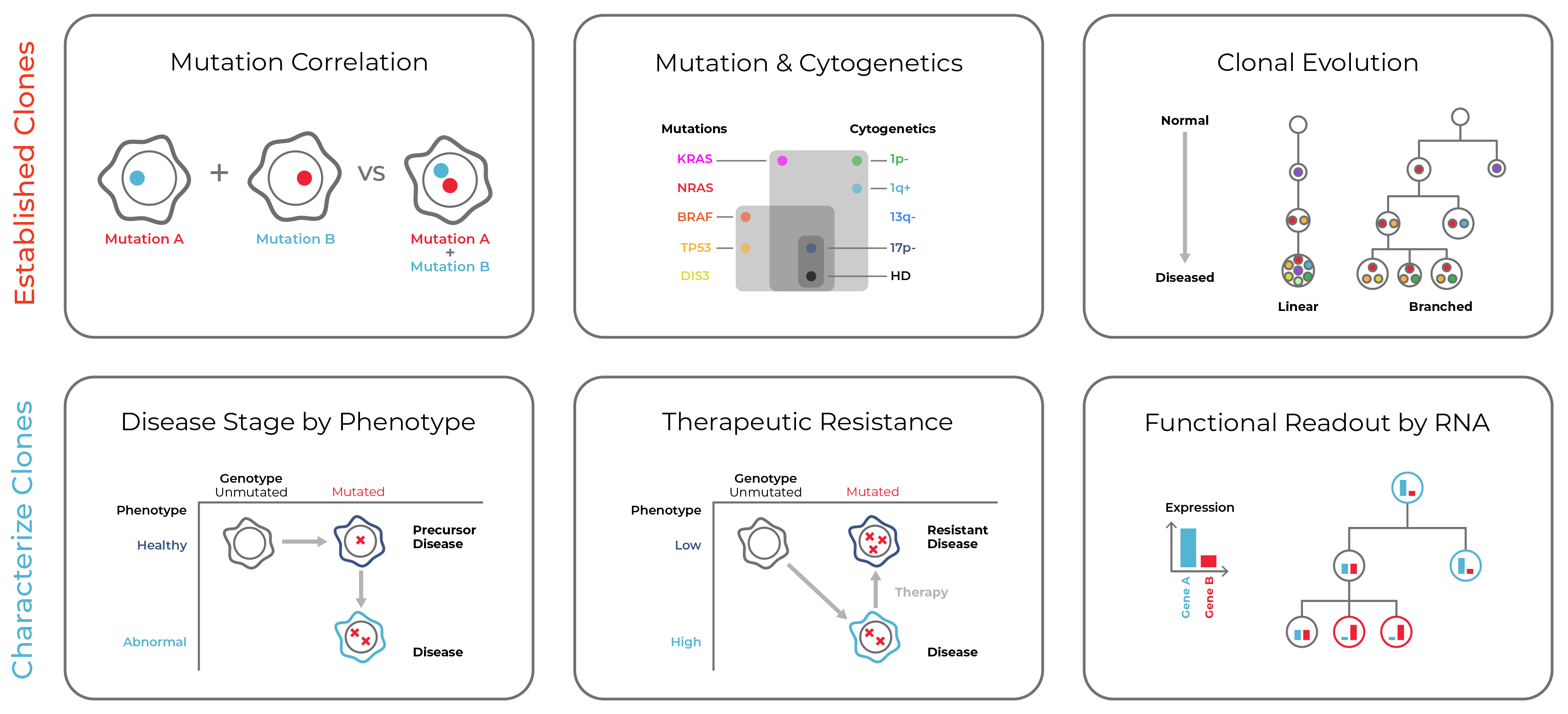
Composite readouts from the Tapestri® single-cell multiomic platform. Top row: established clone identification. Bottom row: clone characterization.
Scoping Your Project
What the Tapestri® Platform Measures
Purpose-built assays that use the Tapestri® single-cell multiomic platform can be configured to measure the following attributes per cell, individually or in combination.
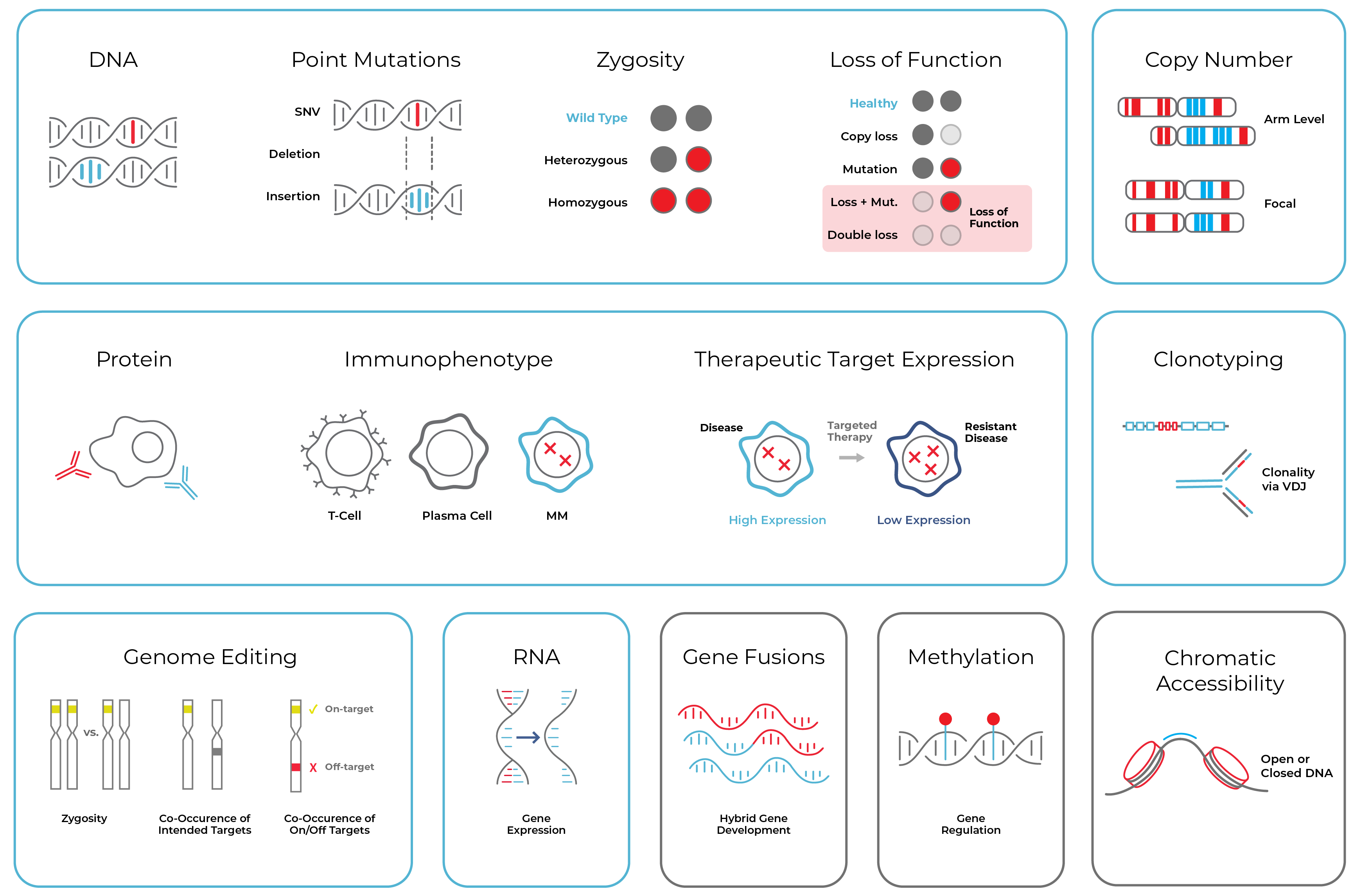
Biological attributes that can be evaluated using Assay Services on the Tapestri® single-cell multiomic platform, including DNA variants (point mutations, zygosity, copy number), protein immunophenotype and therapeutic target expression, genome editing co-occurrence, RNA gene expression, gene fusions, methylation, and chromatin accessibility.
Applications
Disease Areas Across Research and Drug Development
Assay Services delivers single-cell multiomics to researchers spanning hematologic oncology, solid tumors, functional screening, neurobiology, cardiovascular disease, metabolic disorders, and many other diverse applications. Each area below reflects how single-cell multiomics can address the specific biological and therapeutic questions of that context.
Hematologic Oncology
Acute Myeloid Leukemia (AML)
AML is genetically heterogeneous: two patients can carry entirely different driver mutations, and within a single patient, multiple genetically distinct clones coexist. Bulk NGS reports population-average VAFs that cannot determine whether co-occurring mutations like FLT3-ITD and IDH2 reside in the same cell or in separate subclones. Single-cell multiomics directly maps clonal architecture, distinguishes CHIP-associated clones from malignant residual disease, and tracks subclonal evolution under therapeutic pressure for MRD monitoring and drug response stratification.
Myeloproliferative Neoplasms (MPNs)
Myeloproliferative neoplasms (MPNs) include polycythemia vera (PV), essential thrombocythemia (ET), and myelofibrosis (MF). Driver mutations in JAK2, CALR, and MPL co-occur with secondary lesions in genes like ASXL1 and EZH2, and the specific combination drives disease phenotype and transformation risk. Single-cell multiomics resolves which cells carry the compound mutant genotype, supporting stratification of progenitor versus differentiated MPN cell populations in drug trials and enabling monitoring of clonal shifts during treatment.
Multiple Myeloma
Multiple myeloma is driven by plasma cell clones carrying copy number alterations, immunoglobulin gene rearrangements, and secondary mutations that evolve under successive lines of therapy. Single-cell multiomics links clonotyping via V(D)J rearrangement to genetic and phenotypic profiling in the same cell. This allows researchers to track which plasma cell clone expands, what surface target expression (such as CD38 or BCMA) it carries, and how the clone responds to therapeutic agents including proteasome inhibitors, immunomodulatory drugs, and targeted antibody therapies.
Other Leukemias
Across acute lymphoblastic leukemia (ALL), chronic lymphocytic leukemia (CLL), chronic myeloid leukemia (CML), and other leukemias, single-cell genotyping and immunophenotyping characterize the clonal hierarchy driving disease, identify co-occurring mutations that modify therapeutic sensitivity, and track the acquisition of resistance. KMT2A-rearranged leukemias, for example, have been studied with single-cell multiomics to understand how menin-inhibitor resistance emerges at the subclonal level.
Clonal Hematopoiesis of Indeterminate Potential (CHIP)
Clonal hematopoiesis of indeterminate potential (CHIP) describes the age-associated expansion of blood cell clones carrying somatic mutations in genes like DNMT3A, TET2, and ASXL1. CHIP mutations persist after cancer therapy and confound bulk DNA-based MRD assays by mimicking residual leukemia. Single-cell co-measurement of genotype and immunophenotype distinguishes CHIP-associated benign ancestral clones from malignant populations, removing a major source of false-positive MRD calls.
Myelodysplastic Syndrome (MDS)
Myelodysplastic syndrome (MDS) is characterized by somatic mutations in splicing factors, epigenetic regulators, and cohesin complex genes that drive ineffective hematopoiesis and increase risk of transformation to AML. Single-cell multiomics resolves which mutant clone is responsible for specific cytopenias, tracks clonal dynamics in patients receiving hypomethylating agents or targeted therapies, and identifies co-mutation patterns that predict transformation risk independent of bulk VAF measurements.
Other Applications
Solid Tumors
Solid Tumor Research
In solid tumors, intratumoral heterogeneity drives therapeutic failure when drug-sensitive clones are eliminated but resistant minority clones expand. Single-cell multiomics profiles the mutational landscape and surface target expression across individual cells from tumor biopsies or disaggregated tissue, identifying the subclone composition that determines response to targeted agents. This supports the characterization of tumor microenvironment interactions and the design of combination strategies targeting distinct subclonal populations.
Functional Screening
Perturbation Screening and Secondary / Tertiary Validation
Genome-wide and arrayed perturbation screens (including Perturb-seq workflows) generate candidate hits that require mechanistic validation at the cellular level. Single-cell multiomics connects each genetic perturbation to its transcriptional and protein output in the same cell, confirming the mechanism of action at single-cell resolution. Secondary and tertiary screening validation using purpose-built assays on the Tapestri® platform enables prioritization of lead compounds based on their per-cell genotypic and phenotypic impact, rather than population-averaged readouts.
Cardiovascular Disease
Cardiovascular Disease
Cardiovascular disease involves complex interactions among hematopoietic cells, circulating immune populations, and vascular tissue. Inflammatory signaling from clonal immune cell populations contributes to atherosclerosis and other cardiovascular pathologies. Clonal hematopoiesis (CH) is one such contributor: age-related somatic mutations in DNMT3A, TET2, and ASXL1 can produce clonally expanded blood cells with pro-inflammatory transcriptional programs that promote vascular inflammation. Single-cell multiomics characterizes these cell populations at per-cell resolution, connecting genotype to immunophenotype and transcriptional state in the same cell. This supports mechanistic research into how specific immune clones drive cardiovascular inflammation and enables population stratification in preventive or therapeutic cardiovascular programs.
Neurological Disease
Neurological and Neurodegenerative Diseases
Neuroinflammation driven by clonal expansions of microglia, CNS-infiltrating immune cells, and peripheral myeloid populations is increasingly recognized in Alzheimer's disease, Parkinson's disease, and other neurodegenerative conditions. Single-cell multiomics characterizes the genotypic and phenotypic state of these cell populations simultaneously, identifying clonal expansions, co-occurring somatic mutations, and immunophenotypic changes that are invisible to bulk methods. This supports research into the neuroimmune mechanisms that accelerate neurodegeneration and identifies cellular targets for immune-modulating therapies.
Metabolic Disease
Metabolic Disease
Adipose tissue macrophages, liver-resident immune cells, and hematopoietic progenitors with disease-associated somatic mutations contribute to the chronic inflammation underlying type 2 diabetes, non-alcoholic steatohepatitis, and metabolic syndrome. Single-cell multiomics resolves the clonal architecture and functional state of these populations in patient samples, connecting genotype to inflammatory gene expression and surface phenotype per cell. This enables identification of specific mutant clones and cell states that drive metabolic dysregulation, supporting target discovery and biomarker development for immune-targeted metabolic disease programs.
Other Disease Areas
Disease Areas Not Listed Above
Single-cell multiomics on the Tapestri® platform is not limited to the disease areas listed above. Custom panels can be designed around the DNA, RNA, fusions, protein, and epigenetics relevant to your specific disease of interest, so access to single-cell resolution does not depend on working in an already-established application area.
Support
End-to-End Scientific Support Across Your Project
From the first conversation to final data delivery, the Assay Services team takes on every step. Each project is staffed with dedicated scientists who handle assay design, wet lab execution, data analysis, and long-term method support so your team can focus on the biology, not the infrastructure.
Scope Project
Panel & Assay Design
Wet Lab
Data Analysis
Long-term Pipeline Support
Assay Transfer and Scale-Up
Step 01
Scope Project
The Mission Bio Assay Services team meets with your team to understand your targets of interest, sample type, program goals, and any regulatory or transfer requirements. This scoping call defines the assay strategy, sets a realistic timeline, and aligns expectations before any wet lab work begins.
Step 02
Panel and Assay Design
We work alongside your team to design your panels for the Tapestri® platform. Whether you bring your own targets or need assistance selecting the right DNA, RNA, protein, or epigenetic markers, we partner with you to finalize the panels for your project. Panel design incorporates sample constraints, coverage requirements, and multi-attribute needs, with your review and sign-off before moving to the bench.
Step 03
Wet Lab Execution
Mission Bio scientists run the assay in their dedicated laboratory using optimized, validated protocols on the Tapestri® platform. Quality checkpoints are built into the workflow at each stage, from single-cell encapsulation through library preparation and sequencing, to ensure data quality before analysis begins.
Step 04
Data Analysis and Reporting
We process the sequencing data to clearly resolve variants, RNA expression, and protein signal at the single-cell level. Analyzed results are delivered with annotated reports and visualizations that connect directly to your biological question, ready for internal review or regulatory submission.
Step 05
Long-Term Project Support
As your program evolves, our team provides ongoing scientific consultation, offering support for follow-up experiments, result interpretation, and the preparation of methods documentation for publications or assay transfer.
Step 06
Assay Transfer and Scale-Up
When your program is ready to move to a partner site, contract research organization (CRO), or contract development and manufacturing organization (CDMO), the team prepares transfer-ready documentation including installation, operational, and performance qualification (IOQ/PQ) protocols, standard operating procedures (SOPs), and validated analysis workflows. Assays are designed from the start with transfer in mind, reducing failure risk when moving between environments or scaling across development phases.
Resources
Technical Content and Program Guides
Explore technical notes, blogs, and brochures relevant to single-cell multiomic assay services for drug development and disease research.



Talk to a Scientist to Scope Your Assay Project
Whether you are characterizing a disease model, building a biomarker endpoint, or validating a lead compound, the Mission Bio scientific team scopes the assay to your program. No instrument purchase. No new bioinformatics team. Results delivered and transferable.
FAQs
Frequently Asked Questions About Single-Cell Assay Services
What is single-cell multiomic assay service for drug development?
A single-cell multiomic assay service provides fully managed execution of single-cell DNA, RNA, protein, fusion, and epigenetics assays on the Tapestri® platform, including panel design, wet lab execution, data analysis, and results delivery. Rather than purchasing instruments and building internal expertise, research and drug development teams work with Mission Bio scientists who run every step. The service generates per-cell readouts that link genetic variants, transcriptional state, and surface protein expression in the same cell, producing data that separate single-modality assays cannot replicate.
How does single-cell multiomics distinguish clonal hematopoiesis (CH) from malignant disease in hematology samples?
Clonal hematopoiesis (CH) describes the age-associated expansion of hematopoietic cells carrying somatic mutations in genes such as DNMT3A, TET2, and ASXL1. These clones are common in older adults and persist after cancer therapy. Bulk sequencing cannot determine whether a detected variant comes from a benign CH clone or a malignant one: it reports a variant allele frequency but not the cell context behind it. Flow cytometry assigns cells to phenotypic populations but cannot link surface marker expression to the underlying genotype in the same cell. Single-cell multiomics co-measures genotype and immunophenotype per cell, allowing direct separation of CH-associated ancestral clones from malignant populations. This is particularly important in post-treatment hematology samples, where CH-related variants can mimic residual disease and generate false-positive MRD calls without per-cell resolution.
What disease areas and sample types does Mission Bio Assay Services support?
Assay Services supports research and drug development programs across hematologic malignancies including AML, MPN, multiple myeloma, MDS, CLL, and other leukemias, as well as solid tumors, functional perturbation screening, cardiovascular disease, neurological and neurodegenerative disease, and metabolic disease. Supported sample types include bone marrow aspirates, peripheral blood mononuclear cells (PBMCs), disaggregated solid tumor tissue, and cell line models. Custom panels are designed to fit the specific gene targets, surface markers, and multi-attribute readout requirements of each program.
Can single-cell assay results be used for regulatory submissions, and does Mission Bio provide assay transfer documentation?
Assays developed through Mission Bio Assay Services are designed from the start with transfer in mind. For programs approaching regulatory submission, the team prepares IOQ/PQ documentation, standard operating procedures (SOPs), and validated analysis workflows that support transfer to a contract research organization (CRO) or contract development and manufacturing organization (CDMO). The single-cell data generated on the Tapestri® platform can support exploratory and clinical biomarker endpoints in investigational new drug (IND) applications and other regulatory submissions, though the platform and assay services are for Research Use Only and are not cleared or approved for diagnostic use.
How does Mission Bio Assay Services handle clonal evolution and drug resistance tracking in longitudinal studies?
Single-cell multiomics on the Tapestri® platform resolves the clonal architecture of patient samples at the level of individual cells, not population averages. When samples are collected across treatment timepoints, each cell is genotyped and immunophenotyped simultaneously, allowing direct tracking of which subclones contract, persist, or expand under therapeutic pressure. This identifies pre-existing resistance mutations even when the resistant clone is a small fraction of the total population, enabling programs to detect emerging resistance before it drives clinical relapse. Mission Bio scientists design the longitudinal assay strategy during the initial scoping phase and support result interpretation across the full project timeline.